Tumark® Vision Atlas
Case 3
A 45 year-old patient with right-axillary lymph node suggestive of metastasis detected in ultrasound
Dr. S. Metz, Institute for Diagnostic and Interventional Radiology at the Technical University of Munich, Klinikum Rechts der Isar, Munich, Germany
Case description
A right-axillary lymph node, suggestive of metastasis, was detected in an ultrasound. Initially, an ultrasound-guided punch biopsy of the lymph node detected cancerous cells. PET-CT and breast MRI for staging followed. Second ultrasound and sonobiopsy of the inside-right of the breast: NST, G3, HR negative; HER2-immunohistochemistry: 3+.
Course of treatment
The primary tumor and right-axillary lymph node clip marked with Tumark Vision. Primary systemic therapy initiated, consisting of 4 x EC followed by 12 x trastuzumab + pertuzumab weekly. Following primary systemic therapy (PST), complete remission (CR) shown in the imaging.
Conclusion
A treatment option analogous to the course of action suggested in the SenTa study would be a suitable option for the case presented. The hospitals “Kliniken Essen-Mitte” (Kümmel S. et al.) have initiated this study, which is a prospective, multi-centric registry on the usage frequency and feasibility of targeted axillary dissection in patients with primary breast cancer and PST, in whom a punch biopsy is initially performed and the clinically suspicious lymph node is marked with a clip. The primary study goal defined is the identification of the surgical detection rate of the target lymph node (TLNB) marked with a clip. In this context, distinguishing of the clip via ultrasound for pre-operative marking is of crucial importance.
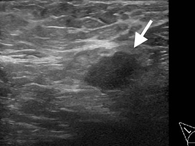
Case 3, fig. 1
The primary lymph node metastasis is indicated by the arrow.
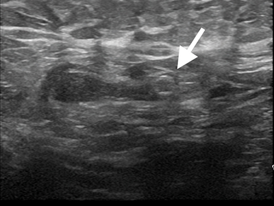
Case 3, fig. 2
Following primary systemic therapy (PST), images show the complete remission (CR) of the metastasis, the lymph node’s inconspicuous morphology, the clip (Tumark Vision) indicated by the arrow, in a regular position, highly visible in the ultrasound.
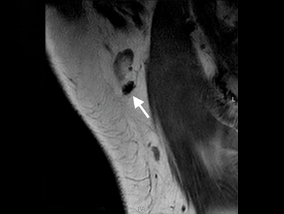
Case 3, fig. 3
MRI corT1-w native to the right axilla; following PST.
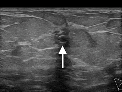
Case 3, fig. 4
Ultrasound of the (former) inside-right of the breast tumor, the clip (Tumark Vision) is clearly distinguished by the arrow. Five months after the initiation of primary systemic therapy (PST).
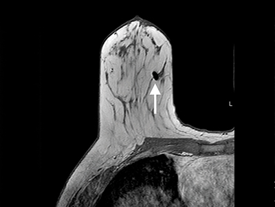
Case 3, fig. 5
MRI axialT1-w of the right breast shows the susceptibility artifact inside due to the clip (Tumark Vision), indicated by the arrow.
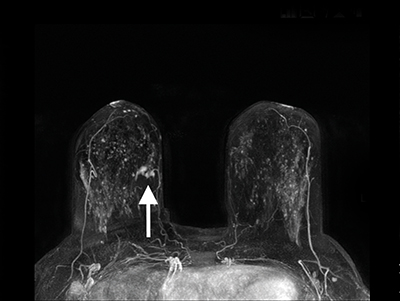
Case 3, fig. 6
MRI as axial maximum intensity projection (MIP) of the first subtraction. Prior to PST with the occult primary tumor inside-right up to this point, is indicated by the arrow.
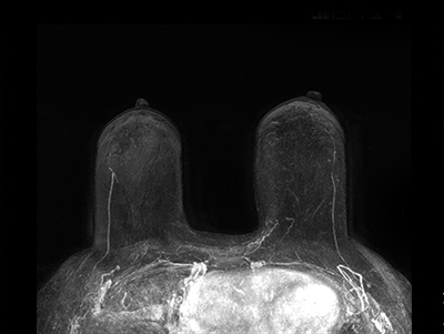
Case 3, fig. 7
MRI as axial MIP of the first subtraction. Image shows CR after PST.